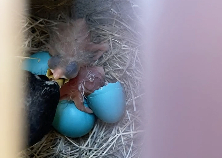
Bird eggs, with their delicate embryos encased in protective shells, have been fine-tuned by millions of years of evolution. In a fertilized egg, each component is optimized to help the chicken embryo grow, protect it from bacterial invasion and predators, and ultimately allow it to break out of the shell and enter the world as a young chick. While the yolk and the egg white are often the stars of this show, especially for lovers of a sunny-side-up fried egg, the thin membrane just inside the eggshell plays an equally important role. It fulfills the dual purpose of providing an additional layer of protection against bacterial invasion and acting as an intermediary between the egg white and the shell.
Marc McKee, professor in the Faculty of Dental Medicine and Oral Health Science and the Faculty of Medicine, recently co-authored a paper with his doctoral student Daniel Buss and Natalie Reznikov, professor in the Department of Bioengineering, zooming in on this membrane, and specifically examining how the wet, organic fibres that make it up attach to the mineral in the eggshell.
“I work in the mineralized tissue field, also known as biomineralization, ” McKee explained in an interview with The Tribune. “It’s a joy for me to work in a field that lies at the intersection of biology, geology, and mineralogy.”
This attachment is intriguing because it binds together a hard inorganic material—the calcium-containing mineral of the shell—and a soft organic material—the fibrous network of the membrane.
Additionally, the bond between them is robust, making it a potentially valuable model for bioengineers looking to adhere other organic fibrous materials to minerals. McKee’s lab set out to investigate how the two materials attach to one another.
“Why is it that when you crack open an egg, you have to peel that membrane off the shell?” McKee said. “And if you look with high-powered electron microscopes, even if you peel it off, and you look at the shell, some fragments of the membranes are still attached.”
The team used X-ray and electron tomography, which involve capturing hundreds to thousands of images that can then be assembled into an incredibly detailed 3D model. Armed with these high-resolution images of the eggshells, they could dive into the detailed structure of the membrane-shell attachment.
First, they confirmed what scientists had already discovered: At the scale of micrometres, some fibres of the membrane actually penetrate the inner portion of the shell. However, using 3D electron microscopy, they could go a step further, zooming in to nanoscale resolution.
“When we delved very deep at the nanoscale into a single fibre using cryo-preservation techniques,” McKee explained, “we discovered that mineral ‘nanospikes’ entered the fibres themselves.”
This ‘nanospiking’ technique is critical because it adds another level of integration, which McKee explained is especially important when you have soft, wet membrane fibres trying to integrate into a hard mineral shell.
“We realized that it’s one thing to incorporate a fibre, but this fibre could still slide in and out of the rock, right? It’s wet, it’s soft, and it’s likely slippery. Within hard eggshell mineral, that attachment ‘rope’ could easily pull out. That’s not a very good attachment, and that would be disastrous for the egg,” McKee said. “We figured out that this nanospiking anchors these wet protein fibres so they can’t move in and out.”
This remarkable mechanism, in which membrane fibres link into mineral at the microscale, and in turn, mineral penetrates into the fibres at the nanoscale, allows for a robust, secure attachment between the two different materials.
While this research is specific to the egg, there are other points of organic-inorganic contact in our bodies, such as where ligaments attach to bones. This discovery presents new insights about the eggshell membrane of bird eggs, and points the way toward further work aimed at understanding what happens when minerals meet organic material.


