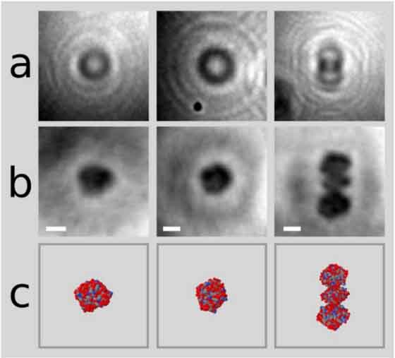
First ever picture of a protein
The study of proteins has always been essential to understanding diseases. Proteins, which are the little worker bees of the human body are responsible for cleaning out debris, transporting vitamins and nutrients, and even fighting off foreign invaders. Because the function of an individual protein is largely dependent on its structure, understanding a malfunction relies on finding mutations in the structure. Scientists have developed a variety of ways to do this, most notably, crystallizing the protein and then taking photos of the crystal using high-energy X-rays, known as X-ray crystallography. Unfortunately, this technique can be time-consuming, costly, and difficult, as it can take months to crystallize a protein. Moreover, exposing proteins to high-energy x-rays can cause damage to the structure.
Consequently, researchers have been looking for alternative imaging methods, and a team of scientists from the University of Zurich have succeeded in finding something that might just work. By using graphene, the carbon allotrope—other allotropes include diamonds and charcoal—the scientists were able to take the first ever photo of a protein without the need of crystals.
“To capture an image of a single protein, the researchers spray a mixture of proteins in solution onto a thin sheet of graphene,” wrote Alexandra Ossola on Popular Science. “They then used a low-energy holography electron microscope, which creates an image by bouncing a beam of electrons off the proteins, then recording how those electrons interact with a pattern of other electrons.”
By using low-energy electrons instead of X-rays, the researchers are able to minimize damage to the protein, and by using graphene as a detector, they are able to obtain a higher level of sensitivity. The researchers took photos of hemoglobin, the protein responsible for carrying oxygen through the body, bovine serum albumin, commonly used in lab experiments, and cytochrome C, a signalling protein. Using this new technology, structural biologists will be able to better understand misfolded proteins in neural diseases like Alzheimer’s and Huntington’s in the hopes of finding a cure.
Reddit AMA used to study rape culture
In July 2012, a thread on the well-known website Reddit sparked some controversy. The comment, titled, “Reddit’s had a few threads about sexual assault victims, but are there any redditors from the other side of the story? What were your motivations? Do you regret it?” was posted on one of their pages—a subreddit—called r/AskReddit. The page is usually used by the Reddit community to answer questions or share funny anecdotes that are normally avoided in regular conversation. This particular question, however, incited a bit of controversy from rape survivors. Due to the anonymity associated with posting on internet forums, many of the posts ended up receiving hateful messages or support; the posts and comments ultimately were all deleted. Before the comments disappeared however, some researchers from Georgia State University were able to get their hands on them. Their goal was to study first-person narratives of sexual assault perpetration.
The work, titled Justifying Sexual Assault: Anoynmous Perpetrators Speak Out Online, took the enormous quantity of comments—over 12,000—from the thread and picked 68 of the first-person accounts to analyze. From their work, they found that many of the perpetrators fell within certain categories: Victim blaming, objectification, biological essentialism, and a hatred of women. Many of the reasons overlapped, but ultimately, the researchers concluded that any and all reason was a self-defence mechanism. By blaming victims or primal needs, the perpetrators do not need to feel shame nor remorse; if they did, then it would mean something was wrong with them and of course that couldn’t be it.
McGill University researchers develop nanoparticle printing press
Nanoparticles continue to gain popularity, and for good reason—they’ve been used in a variety of fields from biomedical supplies and electronics to cosmetics and textiles. Consequently, optimizing their output is vital to furthering the use of nanoparticles across applications. To do this, researchers from McGill’s Sleiman Lab designed DNA nanostructures that acted as chemical ‘sticky patches.’ The nanoparticle is introduced to the sticky patch, where it stays until the researchers dissolve the DNA nanostructure, thereby leaving behind a DNA imprint on the nanoparticle. This DNA-imprinted gold nanoparticle can interact with other DNA-imprinted gold nanoparticles to form specific well-defined assemblies. Because the scientists create the DNA imprint, they have control over the ultimate pattern.
In the past, creating nanoparticle assemblies was difficult because researchers had no way to manipulate or control how the particles would come together. By using a DNA imprint, scientists can create a reproducible and predictable structure. The whole process, the researchers explain, is like going from binding books by hand to having a printing press. The next step, presumably, is seeing what else they will publish.


This is great, will it be a regular feature? It would be great to continue spotlighting lesser-known research at McGill as well!
This is great, will it be a regular feature? It would be cool to continue spotlighting lesser-known research at McGill as well!