
The Maude Abbott Medical Museum is one of the hidden gems of McGill’s downtown campus. Chock-full of real anatomical specimens, tools from bygone eras of medicine, and unnerving 20th-century medical models, the collection is not for the faint of heart, but if you have a strong stomach, it’s worth the trip to the Strathcona Anatomy and Dentistry building to check it out. Here are six items to make sure you see on your visit!
Aviation medicine research helmet
This helmet and control box were part of research carried out by Dr. Geoffrey Melvill-Jones, a physiologist who taught at both McGill and the University of Calgary during his career, and served as a long-time director of McGill’s Aerospace Medical Research Unit (AMRU). During the 1960s and 70s, the AMRU was also involved in early in-orbit experiments and helped to train NASA astronauts to perform experiments during their flights. This particular experiment, which was conducted in 1962, focused on nystagmus—a condition involving uncontrolled eye movements—among pilots.
Miner’s lung specimens
There are actually two exhibits showing the effect of prolonged exposure to mining conditions in the museum. One displays an actual miner’s lung, preserved in 1910, belonging to a miner who suffered from anthracosis—more commonly known as black lung disease. The other is a series of extremely thin slices of miners’ lungs, collected during the 1950s, which are just four micrometres thick and are mounted to filter paper. These cross-sections show the effects of anthracosis and silicosis—a long-term lung disease caused by inhaling fine dust containing silica over many years.
Wax model of vessels and nerves on skull surface
Produced by Maison Tramond, a Paris-based workshop that created anatomical models from wax and bone during the late 19th and early 20th centuries, this skull is one of several jarringly realistic wax models housed in the museum collections. At the time, medical schools were beginning to transition away from using only real specimens and towards more anatomical models, which could be specially produced to illustrate specific maladies and were easier to store and manipulate than organic tissue. This model in particular illustrates what’s going on underneath the skin, highlighting the role of blood vessels and the nervous system.
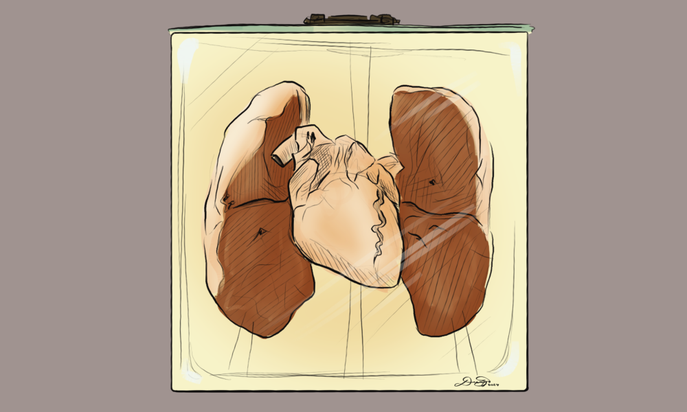
Aspirated peanut
One of many organs showing medical anomalies and fatal conditions in the museum collections, this preserved specimen shows the trachea and lungs of a young child who breathed in a peanut, which became caught in the airway and ultimately caused their death. This specimen, although morbid, is a well-known holding from the collection and a good illustration of the choking hazard posed by having small objects accessible to young children.
Anatomy dissecting lab logbook
Tucked to the side of the museum, this logbook lists the actual names and descriptions of the people whose bodies were donated to McGill’s dissecting lab in the 19th century. The patients cataloged had died of complications from tuberculosis in May of 1896. It goes on to list their ages, religion, and the cemetery they were buried in, all in curly Victorian longhand.
Bonus: Nosce Te Ipsum exhibits
Before you even reach the museum, you may notice that there are several mini-exhibits dotted around the Strathcona Anatomy and Dentistry building, housed inside old fire-extinguisher boxes. It’s hard to miss their bright-red casing, but if you take a closer look, you’ll see that they contain skeletal models, historical drawings of human anatomy, and QR codes if you’re interested in learning more. Each case also sports the motto Nosce Te Ipsum, which means “know yourself” in Latin.