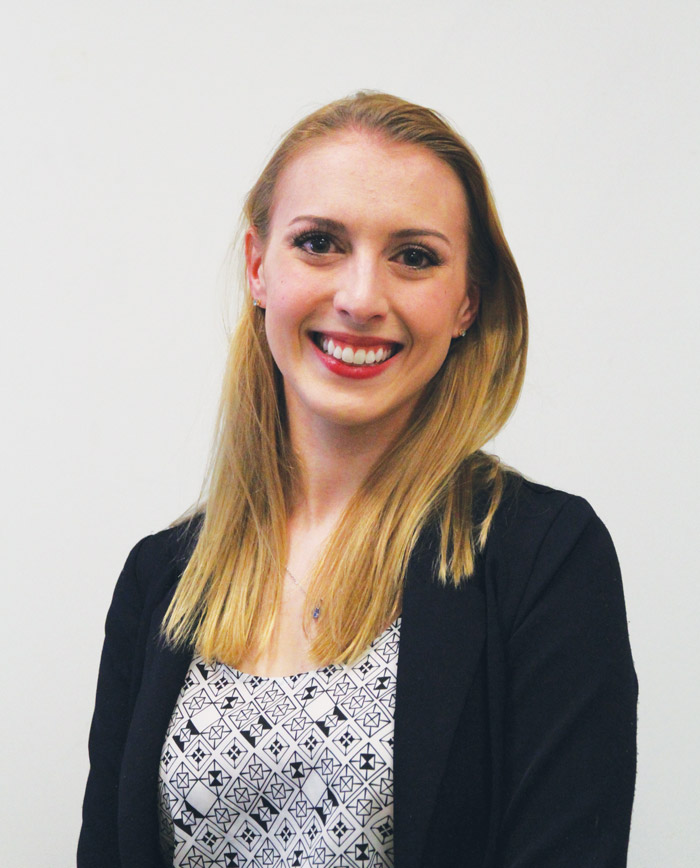
When Valérie Losier, a U3 Physics major, holds up the project she’s been working on for the past academic year, it doesn’t look like the next generation of breast cancer detection technology. Nonetheless, the device—a labyrinth of wires connecting computers and sensors to a bra—may soon become common sight in hospitals and doctors’ offices.
The Popovich lab where Losier has spent the past seven months working is exploring a method of breast cancer detection that moves away from traditional x-ray mammography, which uses the densities of different types of tissues to make a diagnosis. Instead, this new technology uses microwaves to look at tissues’ dielectric properties—a measure of a substance’s ability to reflect and refract radiation.
“The whole basis of this research is that tissues of the breast have different dielectric properties,” Losier said. “You basically shine light [at the tissues], and depending on what is scattered off, you can construct a map [using] the dielectric properties.”
Since the tumours have different dielectric properties, it makes detecting them easy. The detection instrument’s safety presents a major advantage over traditional mammograms.
“X-ray mammography uses X-rays, which ionizes the tissues, and that limits the scans to one time per year, and that’s obviously not optimal,” Losier explained. “This method would be doable once a month because it [uses] microwaves, and they operate at low power [like] wifi and cellphones.”
Because no research has shown any problems associated with extended exposure to these types of waves, the procedure itself is essentially harmless. Losier’s work focuses specifically on the physics of sending out these microwave signals. Essentially, the machine generates signals in the form of wave pulses, which are then read by antennas. Differences in wave readings can then be interpreted to create a map.
“You get 240 antenna-pair readings, [which involve] a lot of algorithms to construct the map,” Losier said. “What I’m involved with is making sure that the signal is sent properly, and that you can read the signal properly in each antenna pair.”
Thus far, the instrument has been tested only in the lab. Losier has been preparing it for clinical trials and anticipates being able to test its accuracy on real tissues awaiting the Glen Hospital relocation project to finish.
Prior to working on breast cancer detection methods, Losier held positions in two other labs.
“I’d done research in physics in my first year,” Losier said. “It was experimental physics working on a satellite instruments [….] Then I did research in atmospheric science after; it was theoretical, numerical work, and then I decided that I wanted to do an engineering master’s and I thought ‘I don’t have experience in engineering’ so I thought I would get involved with a group […] in the engineering department.”
Losier got involved with the lab through a physic’s project course, and strongly recommends using lab courses such as a 396 course—known also as an independent research project—as an introduction to research.
“I just sent [my supervisor] an email,” Losier said. “I’d say [my] advice for anyone is to just get involved in September. Do a 396 course […] and if you do a good job, [the professor] will offer to pay you the next term.”


